
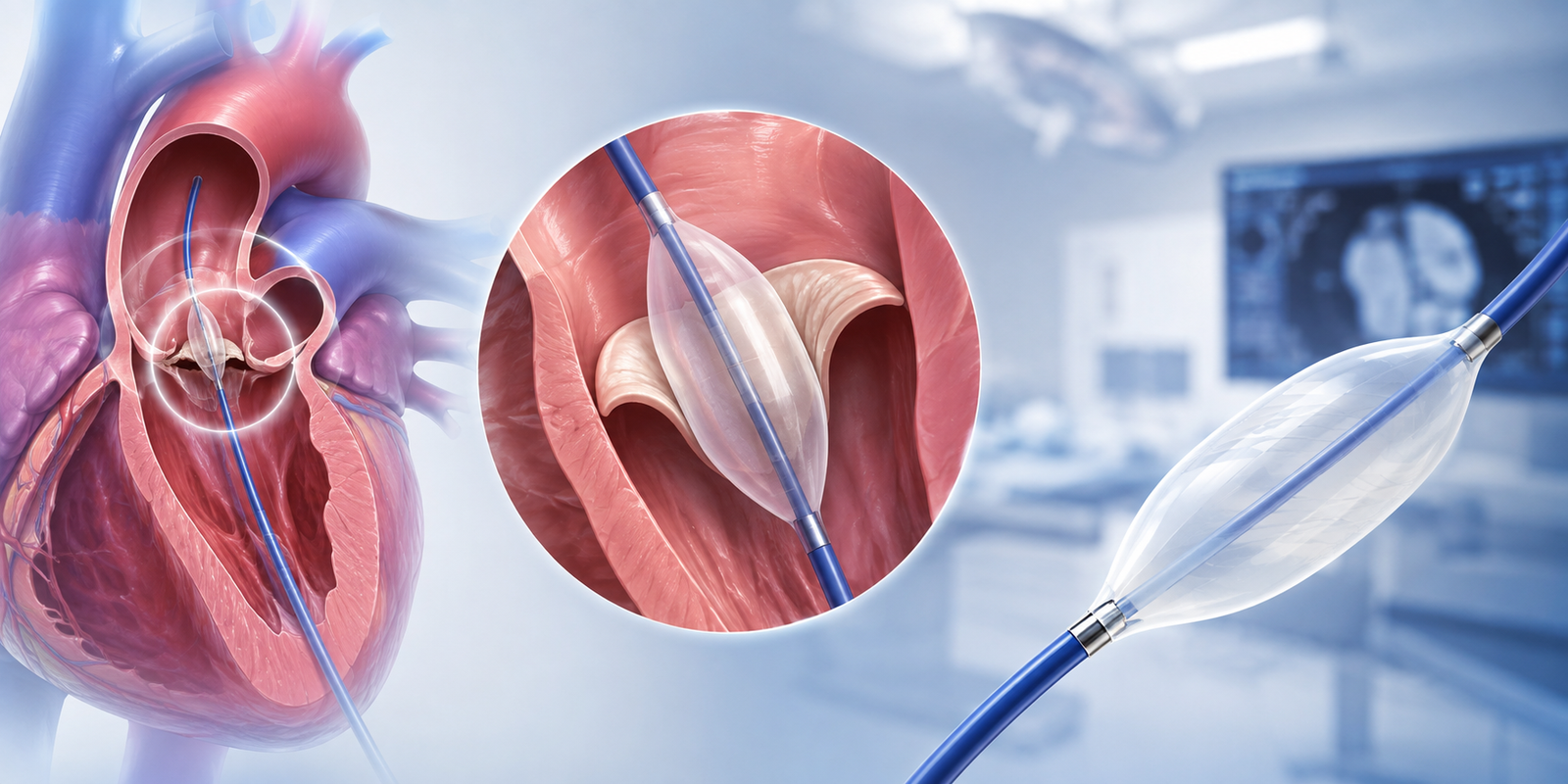
Balloon Valvuloplasty is a minimally invasive heart procedure used to open a narrowed heart valve and improve blood flow. During the procedure, a thin catheter with a small balloon at its tip is guided through a blood vessel to the affected valve. The balloon is then inflated to widen the narrowed valve opening, allowing blood to flow more freely through the heart. It is most commonly used for pulmonary or mitral valve stenosis and, in selected patients, for aortic valve stenosis.
Patients with Narrowed Heart Valves
Individuals with moderate to severe valve stenosis that restricts normal blood flow through the heart.
Patients with Significant Valve Symptoms
People experiencing symptoms such as breathlessness, chest discomfort, fatigue, dizziness, or reduced exercise capacity due to valve narrowing.
Patients Requiring a Minimally Invasive Option
Individuals who may benefit from catheter-based valve treatment because open-heart surgery carries increased risks or is not currently the preferred treatment.
Comprehensive Valve Assessment
Your cardiologist performs an echocardiogram, ECG, blood tests, and other cardiac imaging to assess the affected valve and determine whether balloon valvuloplasty is appropriate.
Catheter & Balloon Placement
A thin catheter is inserted through a blood vessel, usually in the groin, and carefully guided to the narrowed heart valve.
Balloon Inflation & Valve Opening
The balloon is positioned across the narrowed valve and briefly inflated to widen the opening. It is then deflated and removed, allowing improved blood flow through the valve.
Need Expert Medical Guidance?
Your Treatment Journey
Before Procedure
- Your cardiologist will review your medical history, symptoms, medications, and previous heart investigations.
- Imaging tests, particularly echocardiography, are used to evaluate the valve and plan the procedure.
- You may be asked to fast and temporarily adjust certain medications before treatment.
During Procedure
- The procedure is generally performed under local anesthesia with sedation.
- The procedure commonly takes 1–2 hours, depending on the valve being treated and the complexity of the case.
- Your heart rhythm, blood pressure, and other vital signs are continuously monitored.
After Procedure
- You will be observed for several hours, and some patients may stay overnight in the hospital.
- Mild bruising or soreness at the catheter insertion site may occur.
- Most patients can gradually return to normal activities within a few days, following their cardiologist's advice.

Get Expert Medical Guidance
Connect with our specialists and receive trusted healthcare support without delays.
Success!
Your consultation request has been submitted.